Writing in the Proceedings of the National Academy of Sciences, physics graduate student Patrick Noerr, working with professors Ajay Gopinathan, and Kinjal Dasbiswas, collaborated with Jose Zamora from the McCloskey lab to show that elastic substrate-mediated mechanical interactions between cells can drive their self-organization into networks.
Many animal cells actively generate mechanical forces while also sensing and responding to changes in their mechanical environment. This may drive cell–cell interactions through their mutual deformations of the surrounding elastic medium. The physics of this is analogous to electric or magnetic dipoles which may interact with each other at a distance. It was first theoretically suggested by Schwarz and Safran in a seminal paper in 2002 that adherent cells in elastic media may be described as active elastic inclusions that interact with each other through mutual deformations of the medium. Subsequent work, including measurement of cell traction forces, verifies this idea of cells as “force dipoles”.
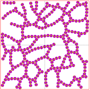
In the present work, Patrick, a graduate student in the Dasbiswas group, took this theoretical picture of “cells as dipoles” and implemented Brownian dynamics simulations of the random movements of cells in the presence of elastic interactions. He used simulations developed by Farnaz Golnaraghi, former graduate student in the Gopinathan group, to show that such dipoles can in principle form networks that resemble structures formed by cells during the development of blood vessels in tissue culture experiments. The physics behind this is quite simple. Dipoles align with each other to form chains, but because of the stochasticity of cell movements (an effective “temperature”), they form networks instead of perfectly ordered chains.
However, does this simple physics work in a complex biological system? To check this, graduate student Jose Zamora cultured human umbilical vein endothelial cells (HUVECs) on soft elastic substrates (a class of materials called “hydrogels”) of varying stiffness. Incidentally, Jose a graduate student in Prof. McCloskey’s lab in the Materials and Biomaterials Science and Engineering (MBSE) department, majored in Physics as an undergraduate here at UC Merced. Jose imaged the cells at several post-seeding times to allow comparison with Patrick’s Brownian dynamics simulations of cells modeled as contractile force dipoles.
We quantified the extent of network formation in both simulations and experiments as a percolation probability order parameter. This is an oft-used statistical physics concept to predict how connected a network is, and whether it can transmit signal across space. We then showed that network formation is maximized at an intermediate substrate stiffness where mechanical interactions are the strongest. Such an optimal stiffness for cell structures was shown before for some other cell types, but this is the first time it was verified in endothelial cell networks. We further predict how network structural features relate to the transport functions of vascular networks and how these may be tuned by manipulating substrate mechanical properties. This understanding of multicellular network formation can guide tissue engineering applications, that require in vitro vascular network formation to be viable.